Diagnostic modalities
Dr. Varun Rai, an ENT and Head & Neck Surgeon at Sir Ganga Ram Hospital in New Delhi, prioritizes patient comfort while delivering precise diagnoses for conditions such as obstructive sialadenitis. This condition frequently manifests as painful gland swelling that worsens during meals, usually resulting from stones or ductal strictures impeding saliva flow.
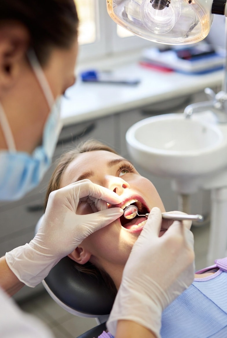
The process typically starts with high-resolution ultrasound, a non-invasive, radiation-free scan that produces real-time images to detect stones, abscesses, or inflammation swiftly and without discomfort.
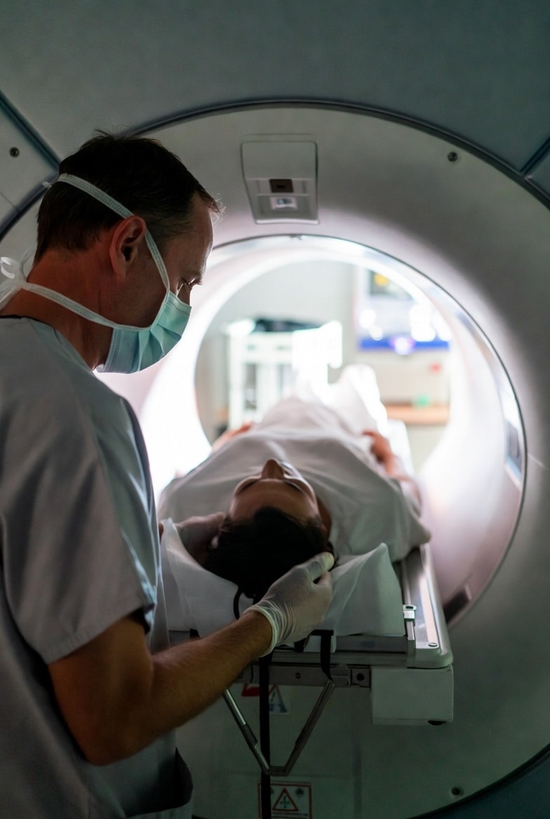
When additional detail is required, MRI sialography provides dye-free, high-resolution imaging of glands and ducts, identifying blockages or abnormalities without needles or X-rays.
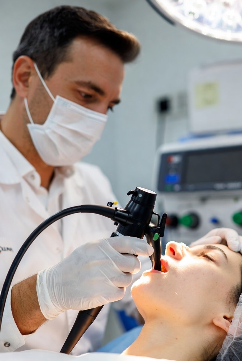
The most advanced option is diagnostic sialendoscopy, involving insertion of a micro-endoscope through the duct's natural opening. This technique enables direct visualization of the obstruction—whether a stone or narrowing—and often facilitates concurrent treatment, delivering immediate relief for many patients upon stone removal.
Dr. Rai tailors the choice of modality to individual symptoms, medical history, and clinical examination, ensuring maximum accuracy with minimal invasiveness. For those experiencing recurrent swelling or pain, prompt evaluation can prevent complications—patients are encouraged to schedule a consultation for personalized assessment.